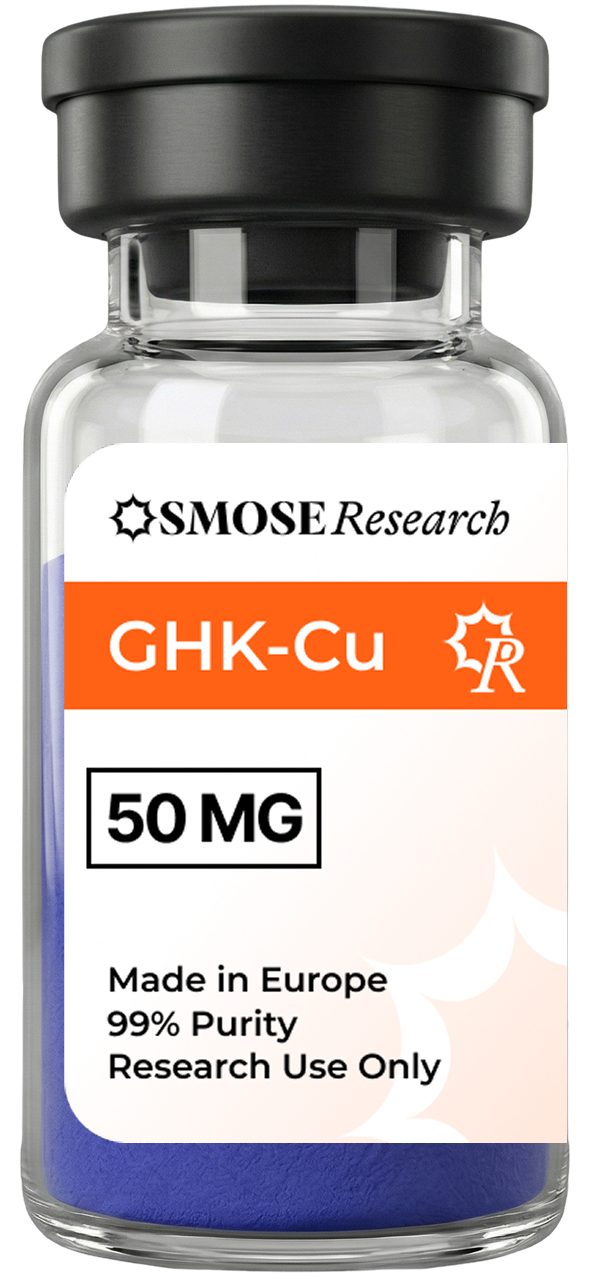
GHK-Cu (Gly-His-Lys-Cu²⁺, 403.93 g/mol) is a naturally occurring copper-binding tripeptide first identified in human plasma by Pickart and Thaler in 1973. Its plasma concentration declines from ~200 ng/mL at age 20 to ~80 ng/mL by age 60, and transcriptomic studies demonstrate modulation of over 4,000 human genes — positioning GHK-Cu as a research tool at the intersection of wound biology, copper biochemistry, and cellular ageing.
GHK-Cu (Gly-His-Lys-Cu²⁺) modulates expression of more than 4,000 human genes in fibroblast models, including COL1A1, COL3A1, SOD2, and integrin β1. Age-related plasma decline (200 → 80 ng/mL) and documented wound-healing acceleration in murine models make it a reference tool in tissue-repair and anti-ageing research. HPLC purity ≥ 99.2% with copper content verification (ICP-MS) required.
The Chemistry of GHK-Cu
GHK-Cu consists of the tripeptide Gly-His-Lys coordinated to one copper(II) ion through a square-planar geometry involving the imidazole ring of histidine (N3), the α-amino group of glycine, and two backbone nitrogen atoms. The binding constant is exceptionally high — log K ≈ 16.44 — resulting in a thermodynamically stable complex under physiological conditions.
The coordination of Cu²⁺ gives GHK-Cu its characteristic bluish-violet colour (d-d absorption at ~540 nm), which serves as a visual indicator of intact complex formation during quality control. Dissociation of copper — for example by chelating agents such as EDTA — produces the colourless apopeptide GHK, which shows markedly reduced biological activity in gene-modulation assays.
The copper delivered by GHK-Cu is functionally relevant: it activates lysyl oxidase (essential for collagen cross-linking), superoxide dismutase (SOD1/2 antioxidant defence), and cytochrome c oxidase (mitochondrial Complex IV) — three enzymatic systems central to tissue homeostasis.
Gene Modulation: Over 4,000 Targets
Pickart and Margolina (2018, DOI: 10.3390/ijms19041987) used gene-array analysis to demonstrate that GHK-Cu modulates the expression of 4,072 human genes in dermal fibroblasts, representing approximately 14% of the annotated human transcriptome. Up-regulated gene sets include:
- COL1A1, COL3A1 (types I and III collagen) — ECM structural proteins
- Integrin β1 (ITGB1) — cell-ECM adhesion and mechanosensing
- Decorin (DCN) — collagen fibril organiser and TGF-β antagonist
- VEGF — angiogenic growth factor
- SOD2, Catalase — mitochondrial and cytoplasmic antioxidant enzymes
Down-regulated gene sets include pro-inflammatory mediators (TNF-α, IL-6) and senescence-associated secretory phenotype (SASP) components. This dual regulation — promoting repair while suppressing inflammation — is the basis for GHK-Cu's research profile in both wound biology and cellular ageing.
Wound Healing and Tissue Repair Models
Fibroblast Models
Campbell et al. (2012, DOI: 10.1155/2012/431024) characterised dose-dependent stimulation of primary human dermal fibroblasts by GHK-Cu, showing increased type I collagen synthesis (70% above control at 10 µM), glycosaminoglycan production (120% above control), and directional migration in scratch-assay models. The effective concentration range is 1 nM to 10 µM, with maximum effects typically observed at 1–10 µM for fibroblast endpoints.
Keratinocyte and Epithelial Models
Park et al. (2009, DOI: 10.1016/j.jdermsci.2009.01.003) demonstrated in HaCaT keratinocyte cultures that GHK-Cu at 1–10 µM increases integrin β1 expression and stimulates dose-dependent directional cell migration — key processes in epithelial wound closure. GHK-Cu treatment also upregulates E-cadherin, supporting epithelial junction re-establishment.
In Vivo Wound Models
Preclinical rodent wound-healing studies document 30–60% reduction in full-thickness wound closure time with topical or subcutaneous GHK-Cu application. Murine incisional models, diabetic wound models (db/db mice), and rat liver-fibrosis models (CCl₄-induced) are the most frequently cited animal systems.
Antioxidant Research
Mas-Bargues et al. (2020, DOI: 10.1016/j.redox.2020.101475) demonstrated that GHK-Cu reduces lipid peroxidation markers by 40% in cellular oxidative stress models and upregulates SOD2 and catalase expression. The mechanism involves copper-mediated activation of SOD and direct radical scavenging via the copper centre. These findings support use of GHK-Cu in research on oxidative-stress-mediated cellular damage, ageing, and metabolic disease models.
Standard In Vitro Models
| Cell model | Application | Typical concentration |
|---|---|---|
| Primary human dermal fibroblasts (HDF) | Collagen I/III synthesis, ECM remodelling | 1 nM – 10 µM |
| HaCaT keratinocytes | Epithelial migration, integrin expression | 1 nM – 10 µM |
| HUVEC endothelial cells | VEGF-driven angiogenesis | 10 nM – 1 µM |
| Mesenchymal stem cells (MSC) | Differentiation, stemness markers | 1 nM – 1 µM |
| 3D skin equivalents (EpiSkin) | Barrier function, stratification | 10 nM – 1 µM |
| Hair follicle explants | Follicle cycling, dermal papilla research | 1–100 nM |
GHK-Cu vs GHK (Apopeptide): Why the Copper Matters
The free tripeptide GHK (without copper) shows substantially reduced gene-modulating activity in transcriptomic studies. The Cu²⁺ ion is required for: activation of lysyl oxidase (collagen cross-linking), SOD activation, cytochrome c oxidase function, and proposed DNA-binding activity at promoter sequences. Researchers comparing GHK and GHK-Cu as matched controls should note that commercially available "GHK" is often a mixture of the apopeptide and variable copper-bound fraction — underlining the importance of ICP-MS copper content verification in quality-controlled GHK-Cu batches.
Quality Requirements
For quantitative gene-expression studies, RP-HPLC purity ≥ 99.2% (peptide component) must be combined with ICP-MS copper content verification to confirm the 1:1 Gly-His-Lys:Cu²⁺ stoichiometry. The colour of the reconstituted solution (pale blue-violet) provides a qualitative indicator, but is not a substitute for analytical data. OSMOSE Research supplies GHK-Cu with batch-specific RP-HPLC chromatogram, ESI-MS mass confirmation (403.93 Da), ICP-MS copper content, and LAL endotoxin test (< 0.5 EU/mg).
Reconstitution
Lyophilised GHK-Cu dissolves in sterile water or PBS pH 7.4. Avoid EDTA-containing buffers (e.g. standard cell culture PBS EDTA formulations) that will chelate and remove the copper ion. Stock concentration typically 1–10 mM; working concentrations 1 nM to 10 µM. The pale blue-violet colour of the solution confirms intact Cu²⁺ coordination. Stable for 72 hours at 4°C; store aliquots at -20°C for longer periods.
FAQ
Why does GHK-Cu modulate so many genes?
GHK-Cu appears to interact with gene promoter sequences — specifically DNA motifs in the major groove — as proposed by Pickart et al. based on structural homology of the His-Cu-Lys coordination complex with known transcription factor metal-binding domains. Additionally, the copper delivered to intracellular compartments activates metalloenzymes (SOD, cytochrome c oxidase, lysyl oxidase) that have broad downstream transcriptional effects through redox signalling.
What is the optimal GHK-Cu concentration for fibroblast studies?
The dose-response profile is typically bell-shaped: maximal effects on collagen synthesis and migration are observed at 1–10 µM, with reduced effects at higher concentrations (> 100 µM) due to potential copper toxicity. For gene-array studies, 1 µM for 24–48 h is the most commonly used condition (Pickart and Margolina, 2018). A four-point dose-response (0.1 nM, 10 nM, 1 µM, 10 µM) is recommended for characterising new endpoints.
Does GHK-Cu affect hair follicle biology?
Preclinical hair-follicle explant studies and scalp fibroblast models have documented upregulation of hair-growth-associated genes (KRT81, KRTAP proteins, growth factors) by GHK-Cu. Specific mechanisms proposed include activation of dermal papilla cells and extension of the anagen (growth) phase. These remain active research questions without conclusive mechanistic clarity.
How is copper content in GHK-Cu verified?
ICP-MS (inductively coupled plasma mass spectrometry) or ICP-OES (optical emission spectrometry) quantifies the copper content in µg/mg of peptide complex. The expected copper content for a 1:1 GHK:Cu²⁺ complex (MW 403.93 g/mol) corresponds to 15.77% w/w copper. Discrepancy from this value indicates partial decomplexation or incorrect stoichiometry. OSMOSE Research includes ICP-MS copper data in the CoA for all GHK-Cu batches.
Can GHK-Cu be used in cosmetic formulation research?
Our GHK-Cu is supplied for in vitro research use only, including cosmetic ingredient research (efficacy testing in cell models, 3D skin equivalents). It is not pre-formulated for finished cosmetic products and does not hold a cosmetic product notification. Researchers developing cosmetic applications must perform their own product safety and regulatory assessments under applicable EU cosmetics regulation.
Disclaimer — Research use only
The information in this article is provided for informational purposes for the scientific community. The products mentioned are intended exclusively for in vitro research and are not approved for human or animal use. Administration to any living being is strictly prohibited. See the legal page.

OSMOSE Research
Research team
European research peptide supplier. Our articles are written from scientific literature published in peer-reviewed journals.
Related articles
Education
Research Peptides Glossary: Definitions of BPC-157, TB-500, GHK-Cu & More
Scientific definitions of the most-researched peptides: BPC-157, TB-500, GHK-Cu, Melanotan-2, CJC-1295, Tesamorelin, Epithalon, MOTS-c, and Semax — with preclinical research context and real PubMed citations.
Research
Melanotan-2 (MT2): Research Guide to Melanocortin Pharmacology
Preclinical research guide to Melanotan-2 (MT2, MT-II, MTII): MC1R–MC5R receptor pharmacology, melanogenesis models, cyclic structure, and EU-grade purity requirements.
Research
TB-500 (Thymosin β-4 Fragment): Research Guide 2026
In-depth preclinical research guide to TB-500: actin-binding mechanism, cellular migration models, cardiac and corneal applications, LKKTETQ sequence data and CoA standards.